
Blog
Elemental Imaging of Biological Samples Using J200 Tandem LA–LIBS
The J200 Tandem LA–LIBS combines LA-ICP-MS and LIBS to map trace and major elements from the same sample — demonstrated on tumor, skin, brain, and leaf tissues.
By Dr. Alexander Bolshakov · Bio
Laser ablation (LA) is a versatile analytical tool for direct solid sampling. A pulsed laser beam is focused onto a sample to remove a small amount of material, either for direct optical analysis of the resulting plasma emission by laser induced breakdown spectroscopy (LIBS), or for transporting the ablated mass to an inductively coupled plasma (ICP) with subsequent detection by mass spectrometry (MS). Applied Spectra's Tandem LA–LIBS instruments perform both measurements simultaneously from the same laser pulse, combining the analytical merits of both techniques, LA-ICP-MS and LIBS.
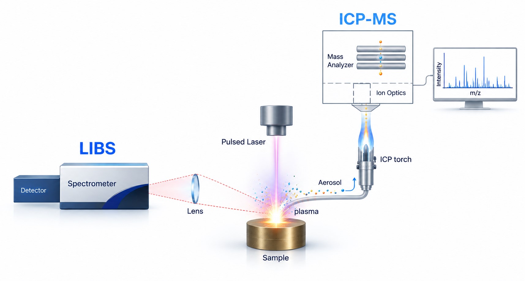
LA-ICP-MS provides highly sensitive detection of trace elements and isotopes, while LIBS detects all elements, including the light elements such as H, Li, Be, B, C, N, O, F, and Na that are difficult or impossible to measure by ICP-MS. Combining both techniques in a single measurement yields a complete elemental picture — trace analytes by LA-ICP-MS and major and light elements by LIBS — from the same sampling location. Because the laser samples the solid directly, imaging by Tandem LA–LIBS may involve only minimal or no sample preparation.
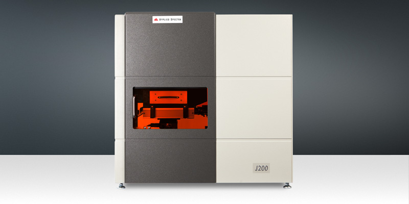
Several examples of using the J200 Tandem LA–LIBS Instrument for bioimaging follow.
Imaging of human pleural mesothelioma
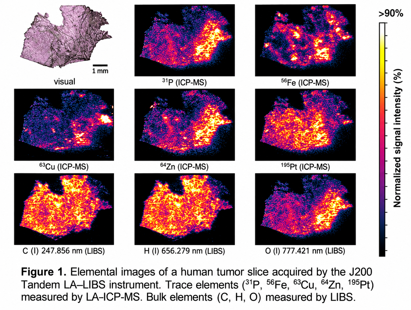
A slice of a human tumor treated with the anticancer drug cisplatin was imaged to reveal the distribution of platinum (Pt), the active metal in cisplatin, together with the endogenous elements phosphorus (P), iron (Fe), copper (Cu), and zinc (Zn) by LA-ICP-MS, and the bulk constituents carbon (C), hydrogen (H), and oxygen (O) by LIBS. The trace analytes and the bulk elements were mapped simultaneously from the same ablation, providing complementary chemical context for the drug distribution within the tissue.
Imaging of porcine whole skin in vitro
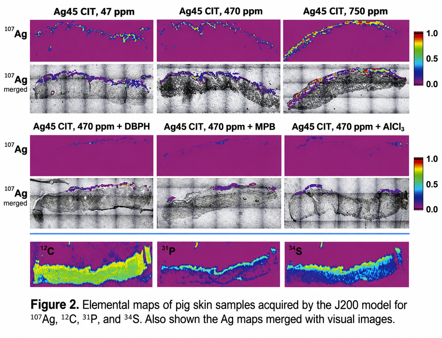
Silver nanoparticles are widely used for their antimicrobial properties, and their penetration through skin is of interest for safety assessment. Whole pig skin exposed to silver nanoparticles at different concentrations was imaged for silver (Ag) by LA-ICP-MS and for carbon (C), phosphorus (P), and sulfur (S) by LIBS. The silver maps were merged with visual images of the tissue to localize the penetration of silver relative to the skin structure. The highest amount of Ag that penetrated into the skin was 0.45 ng/cm² as measured for the highest exposure concentration.
Imaging of rat brain in vitro
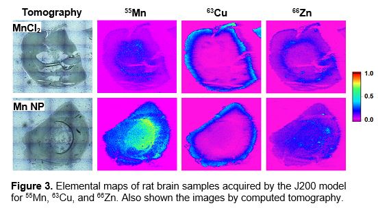
Manganese is an essential trace element, and manganese-based nanoparticles are being investigated as contrast agents. Rat brains were treated with either a manganese chloride (MnCl₂) solution or manganese nanoparticles (Mn NP), and thin brain sections were imaged for manganese (Mn), copper (Cu), and zinc (Zn). The elemental maps reveal the distribution of manganese across the brain structures, with computed tomography images shown alongside for anatomical reference.
Mapping of contaminated leaves
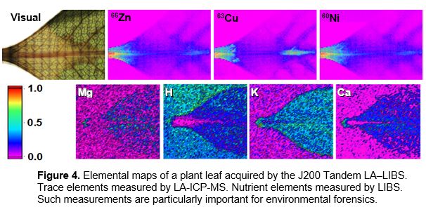
Plant leaves exposed to metal contamination were analyzed to map the uptake and distribution of the contaminant elements across the leaf surface. Direct solid sampling by laser ablation allows the leaf to be imaged with little sample preparation, preserving the spatial distribution of the elements as they occur in the tissue. The resulting elemental images are shown in Figure 4.
Femtosecond LA-ICP-MS and LIBS imaging
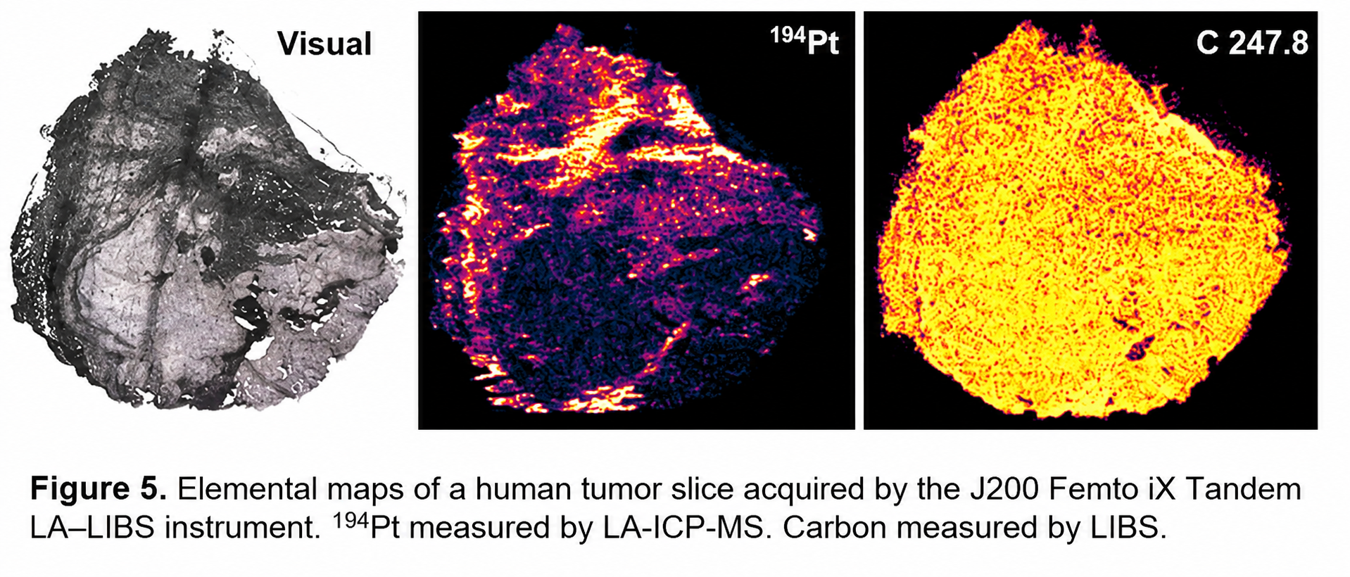
Using the J200 Femto iX Tandem LA–LIBS instrument, a human tumor slice was imaged with femtosecond laser pulses. Platinum (¹⁹⁴Pt) was measured by LA-ICP-MS and carbon by LIBS. Femtosecond ablation produces well-defined ablation craters with minimal thermal effects, improving spatial resolution and image quality for the elemental maps.
These examples demonstrate that the J200 Tandem LA–LIBS instrument provides simultaneous, spatially resolved imaging of both trace and major elements in biological samples, delivering a more complete chemical understanding of tissues from a single measurement.